

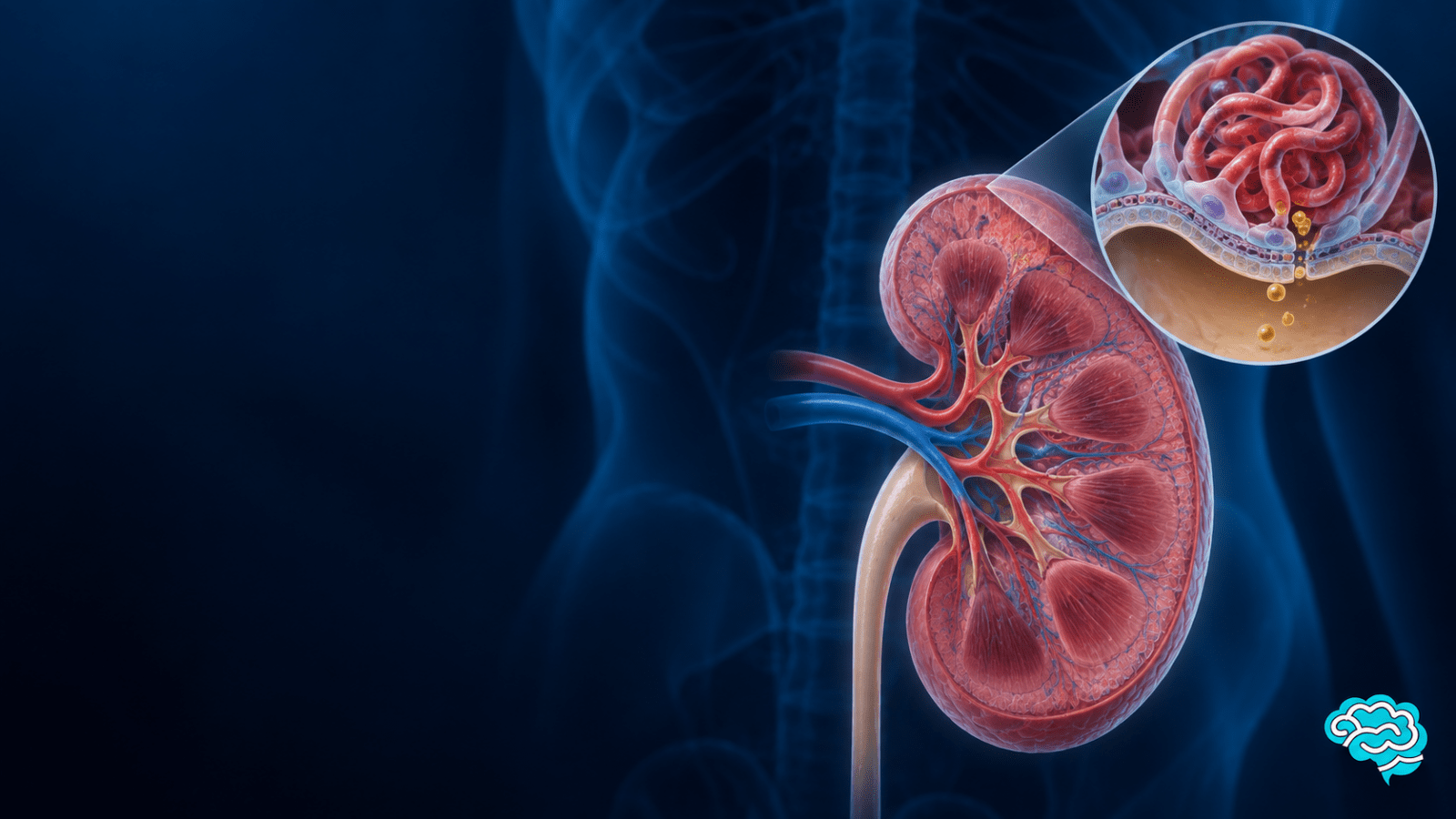

Nephrotic syndrome is usually recognized by a patient with edema and significant protein loss in the urine. However, memorizing these findings does not explain why they occur together. In this article, we address the pathophysiology of nephrotic syndrome, first we will see how the glomerular filter works and then we will follow the pathway from its injury to the clinical signs.
What is nephrotic syndrome?
Nephrotic syndrome is a set of manifestations produced by a disturbance of the filtration barrier of the glomerulus..
Its central feature is the intense proteinuria: an abnormal amount of proteins, especially albumin, passes through the renal filter and is eliminated in the urine. When albumin is lost, its concentration in the blood decreases; this is called hypoalbuminemia.
Edema and hyperlipidemia complete the classic picture, although they do not have to appear with the same intensity in all patients.
In adults, the threshold of 3,5 g of protein in urine over 24 hours, or a urinary protein/creatinine ratio close to 3,5 g/g, is commonly used to refer to nephrotic range proteinuria.
In children, the KDIGO guideline 2025 defines this range by a protein/creatinine ratio of at least 2 g/gamong other criteria adjusted for age and body surface area. It is important to keep this in mind and not make the mistake of automatically applying the same number to both groups.
NOTE: The nephrotic range proteinuria (more than 3.5g of protein in urine over 24 hours) does not by itself equate to complete nephrotic syndrome. This is because there are other etiologies that can cause protein loss in urine.
The diagnosis of nephrotic syndrome must include, in addition to proteinuria; urinary loss with serum albumin and the clinical situation. This distinction prevents confusing a laboratory result with the entire pathophysiological process.
Renal anatomy
The kidney contains nephrons, its working units. Each nephron begins in a glomerulus, a tuft of capillaries to which blood arrives via the afferent arteriole and exits through the efferent arteriole.
The fluid that crosses its walls enters the Bowman capsule and continues through the tubules; there, water and useful substances are recovered before forming the final urine.
Remember that the structural and functional unit of the kidney is the nephron.
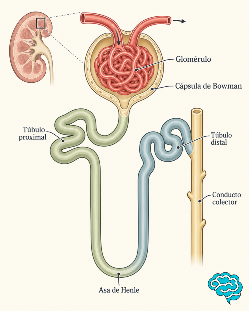
Image 1 – Renal Anatomy
As we explained in our article on glomerular anatomy and physiology, the filter is not a single wall.
It has three components that work together: the capillary endothelium with its glycocalyx surface, the glomerular basement membrane, and the podocytes, whose extensions form the slit diaphragms.
If it helps to visualize, think of a multi-layered filter; but remember that here cellular proteins and physical forces are also involved, which a simple mesh does not represent.
The podocytes are especially important. Their foot processes embrace the capillary and leave slits regulated by proteins such as nephrin.
An injury to their foot processes, the basement membrane, or other components of the filter can change the permeability to proteins.
Normal renal physiology
The glomerulus filters large amounts of water and small solutes, but restricts the passage of cells and most plasma proteins.
Albumin, due to its size and the properties of the entire glomerular barrier, crosses only in small amounts..
The fraction that reaches the proximal tubule can be recovered; however, that capacity has a limit. Filtration depends on the pressure in the glomerular capillaries and the forces opposing the outflow of fluid.
The tubules, for their part, regulate how much sodium and water are returned to circulation. Thus, to maintain balance, it is not enough to filter: it is also necessary to decide what is reabsorbed. This second function will be fundamental in explaining edema.
Circulating albumin contributes to the oncotic pressure, the force with which proteins help keep water within the vessels.
Here it is worth making a small reminder about osmosis. Remember that water can cross the capillary wall and tends to move toward areas where there is a higher concentration of particles that cannot cross it. In blood vessels, albumin is the main protein responsible for this effect, favoring that water remains within the intravascular space.
When the kidney loses too much albumin, it changes that balance. The liver tries to compensate by increasing protein production, but sometimes it is not enough to match urinary loss.
Causes of nephrotic syndrome
Nephrotic syndrome describes what happens, not the name of a single disease.
Always remember that when we talk about “SYNDROME” we are not actually talking about a disease itself but rather a set of diseases or pathologies that share signs or symptoms.
A useful way to organize its causes is to ask whether the problem originates mainly in the glomerulus or if a systemic disease is damaging it.
Primary glomerular diseases
Among the primary causes, the minimal change disease stands out., the focal segmental glomerulosclerosis (FSGS) and the membranous nephropathy.
In minimal change disease, optical microscopy may show few findings, while electron microscopy reveals podocyte effacement. It is a common cause in children with steroid-sensitive nephrotic syndrome.
FSGS describes scars in some glomeruli and in a portion of each affected glomerulus. It can be primary, secondary to another insult, or of genetic origin; this classification matters because not all variants are treated with immunosuppression.
In membranous nephropathy, immune deposits damage the subepithelial area of the barrier. In some cases, antibodies against the phospholipase A2 receptor, known as PLA2R, are detected.
Secondary causes of nephrotic syndrome
Diabetes, systemic lupus erythematosus, and amyloidosis can cause intense proteinuria and nephrotic syndrome.
We should also consider infections such as hepatitis B, hepatitis C, and HIV; some medications, including non-steroidal anti-inflammatory drugs; neoplasms in selected contexts; and genetic diseases affecting podocyte proteins. Age, history, progression, and extrarenal findings guide the search.
This list explains an essential clinical decision: before saying “corticosteroids should be given,” we must know what is damaging the glomerulus. Treating a primary podocytopathy, diabetic nephropathy, and an active infection in the same way would ignore the cause of the syndrome.
For example:
- Hepatitis B, hepatitis C, and HIV:can cause secondary glomerular diseases. Here, it is not enough to immunosuppress; the infection must also be treated or controlled.
- NSAIDs(ibuprofen, naproxen, diclofenac, etc.) can be associated with some glomerular diseases, especiallyminimal change diseaseand, occasionally, membranous nephropathy. In this context, a fundamental measure isto discontinue the causative medication..
- Neoplasms:some cancers may be associated with glomerular diseases. For example, certain solid tumors are related tomembranous nephropathy, while Hodgkin lymphoma may be associated withminimal changes.
- Genetic diseases:mutations in proteins important for the podocyte — such asnephrin or podocin— can alter the filtration barrier and produce proteinuria. In these cases, corticosteroids may have little or no utility.
Therefore, age and history are important. A previously healthy child with nephrotic syndrome has likely causes different from those of a 65-year-old adult with diabetes, weight loss, or hepatitis C.
Epidemiology
Nephrotic syndrome occurs in children and adults, but its frequency and causes change with age and the studied population.
In a cohort of adults from Denmark, the recorded incidence ranged from 3,35 to 4,30 cases per 100 000 person-years between 1995–2000 and 2013–2018. This is an estimate from that health system; it does not represent a universal rate nor does it allow for the assumption of the same frequency in Latin America.
In childhood, KDIGO reports a wide variation between populations, of approximately 1,15 to 16,9 cases per 100 000 children.
Minimal change disease is closely linked to steroid-sensitive nephrotic syndrome in children.In adults, the causes are distributed differently.
Signs and symptoms of nephrotic syndrome
The edema is usually the most visible manifestation. It may begin around the eyes, become evident in the ankles and legs, or extend to the abdomen and other areas.
Rapid weight gain may reflect fluid retention, not necessarily an increase in adipose tissue.
In severe cases, there may be ascites, pleural effusion, or generalized edema, known as anasarca.
Urine may appear foamy due to the abundance of proteins, although isolated foam does not confirm a diagnosis.
The reason for the foam is physical: proteins have propertiessurfactant, meaning they reduce the surface tension of urine. When urine hits the water and traps air, bubbles are formed; proteins help tostabilize them, making the foam more abundant and lasting longer. Essentially, a phenomenon similar to what we see when soap facilitates foam formation occurs.
There may also be fatigue, decreased appetite, and a feeling of heaviness. Some patients present with an abnormal urine analysis before developing noticeable edema.
We must look for complications in addition to the initial symptoms: venous thrombosis, infections, acute kidney injury, and progressive deterioration of renal function.
Apainful leg that is more swollen than the othershould raise suspicion of possible deep vein thrombosis, especially since nephrotic syndrome promotes a state of hypercoagulability.
Similarly, sudden respiratory difficulty, fever, or a marked decrease in diuresis require prompt clinical evaluation, as they may indicate complications such as pulmonary embolism, infection, or acute kidney injury, beyond the usual edema of nephrotic syndrome.
Pathophysiology of nephrotic syndrome
Glomerular injury in nephrotic syndrome
Let us remember what we learned at the beginning of this article. The glomerular barrier restricts the passage of proteins. When podocytes, the basement membrane, or their interactions are damaged, albumin crosses in an amount that exceeds the tubular recovery capacity.
The result is persistent proteinuria. The specific injury varies according to the disease: there is no single molecular pathway that explains all nephrotic syndromes.
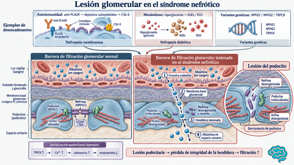
Image 2 – Glomerular injury in nephrotic syndrome
There are different causes that can damage the glomerular filtration barrier. In the image 2 at the top, three examples are presented: in membranous nephropathy, anti-PLA2R antibodies bind to a podocyte antigen and promote the formation of immune deposits and complement activation
In diabetic nephropathy, hyperglycemia is associated with advanced glycation end products and oxidative stress; and, in some hereditary forms, variants ofNPHS1 (Nephrin), NPHS2 (Podocin) or TRPC6alter important proteins for podocyte function.
These are different causes that can lead to similar injury.
On the left in the Image 2, the normal barrier is represented: blood albumin successively encounters the endothelium and its glycocalyx, the glomerular basement membrane, and the podocyte foot processes. Between the latter is the slit diaphragm, in which nephrin and podocin participate, connected by proteins such as CD2AP and Nck to the actin filaments that support the shape of the foot process.
Damage in Nephrotic Syndrome is found in most cases at the level of the Podocytes. However, some pathologies can also cause damage at the level of the glomerular basement membrane and the endothelium.
To the right of the image 2 we can observe the molecular process of the pathophysiology of nephrotic syndrome, nephrin and podocin appear disorganized, actin loses its normal arrangement and the foot processes widen and flatten, a phenomenon calledfoot process effacement.
Nephrin and podocin are essential for maintaining the slit diaphragm between the foot processes of the podocytes.In practical terms, they are like a toll booth that is part of the glomerular filtration barrier. If these proteins fail, the structure of the slit may be altered and the barrier allows a greater amount of proteins, especially albumin, to pass into the urine.
It is important to mention that the mechanism of injury to the podocyte and/or glomerular basement membrane and endothelium depends on the disease causing it.
In thegenetic formsthe alteration may begin in the protein itself. For example, some variants ofNPHS2 cause podocin to be retained within the cell; as it interacts with nephrin, it may also prevent nephrin from reaching the membrane properly.
Indiabeteshyperglycemia can modify podocyte signaling and promote the removal of nephrin from the cell surface; in biopsies of diabetic nephropathy, lower expression of nephrin, podocin, and synaptopodin has also been observed.
Inmembranous nephropathy associated with anti-PLA2R, damage may begin with the binding of the antibody and the activation of the complement. Damaging and displacing NEPH1 (Nephrin) and synaptopodin or Nephrin, Podocin
The TRPC6 → Ca²⁺ → calcineurin → decrease in synaptopodin pathway illustrates one of the mechanisms that can contribute to this disorganization, but it does not necessarily occur in all cases.
When synaptopodin decreases, it also affects the actin of podocytes. This is because synaptopodin helps to support the architecture of actin.
Finally, the dotted line shows the path of albumin from the capillary, through the damaged barrier and the altered slit, to the urinary space;it does not pass through the interior of the podocyte. Thus, the common result of these injuries is the increased filtration of albumin and its loss in the urine.
Why does a microscopic alteration produce a sign that we can see with the naked eye? Because the glomerulus continuously filters blood. A small but sustained leak of an abundant protein can add up to grams per day; when that loss exceeds replacement, serum albumin decreases. This is the first connection between kidney injury and systemic manifestation.
Consequences of albumin loss
With less circulating albumin, one of the forces that keeps fluid within the vascular space decreases. Part of the water moves to the interstitium, where edema appears.
If the effective circulating volume falls, the body may interpret that there is a lack of fluid even though the patient is “swollen.”
As a consequence of this misinterpretation, mechanisms for sodium and water retention are activated, including the renin-angiotensin-aldosterone system.
This model is often referred to as underfill.
But edema is not always explained by that sequence. Some patients have normal or increased plasma volume. Therefore, assuming that every edematous person is intravascularly ’empty’ can lead to incorrect decisions.
Clinical examination, blood pressure, renal function, and response to treatment help interpret each case.
Renal sodium retention
There is another pathway: the kidney inappropriately reabsorbs sodium, and water follows it. When the glomerular barrier allows proteins that normally do not reach the tubule to pass, some may participate in the activation of the epithelial sodium channel (ENaC) in distal segments.
Experimental and observational studies relate urinary proteins such as plasminogen and plasmin to that activation. This mechanism helps explain edema even without an initial reduction in circulating volume.
This model is often referred to as overfill.
In practice, underfill and overfill are models to understand mechanisms that may coexist.
The important thing is the complete chain: glomerular injury → protein loss → changes in vascular forces and renal sodium reabsorption → accumulation of water in tissues. It is not necessary to choose a single model for all patients.
Underfill vs Overfill
| Underfill | Overfill |
|---|---|
| First ↓ oncotic pressure | First ↑ renal retention of Na⁺ |
| Water leaves the vessels | Kidney retains Na⁺ and water |
| ↓ effective circulating volume | Normal or increased circulating volume |
| RAAS is activated | Retention can occur without activating RAAS |
| Retention issecondary/compensatory | Retention isprimary renal |
NOTE: as we mentioned, these models they are not necessarily exclusive mechanisms. A patient with nephrotic syndrome may present components of both, and which predominates depends on the cause and the stage of the disease.
A very simple way to remember it would be:
UNDERfill:…“water spills out of the glass → the kidney tries to recover it.”
OVERfill:Nephrotic syndrome“the kidney retains too much salt and water → it overflows into the tissues.”
Hepatic Response to Nephrotic Syndrome
In response to the drop in albumin, the liver increases protein synthesis.
In this context, the production and metabolism of lipoproteins are also altered; therefore, cholesterol and triglycerides may increase.
Additionally, some lipid particles appear in the urine and cause lipiduria, which can be observed as fatty casts or fat bodies in the sediment. The intensity of hyperlipidemia varies and usually improves when the syndrome resolves.
The Lipiduria is the presence of fats or lipids in the urine.
It may appear in the urine as:
- Fat droplets
- Oval fat bodies
- Fatty casts
When observed under polarized light, some lipids produce the characteristic image of“Malta cross”.
Effect of hypoalbuminemia on coagulation and immunoglobulins
Urine does not only carry albumin. The loss of immunoglobulins and other defense components, along with the effects of certain treatments, increases vulnerability to infections.
In parallel, the loss of proteins that inhibit coagulation and the hepatic response that increases procoagulant factors favor a state of hypercoagulability.
The risk of thrombosis is not identical for everyone. It depends, among other factors, on the underlying disease, the degree of hypoalbuminemia, immobility, and personal history.
In a cohort of membranous nephropathy, low albumin was associated with a higher risk of venous thromboembolism; this data supports monitoring, but does not mean that every patient should receive preventive anticoagulation.
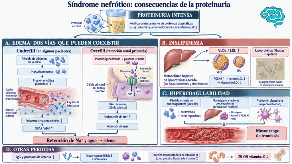
Image 3 – Consequences of proteinuria in nephrotic syndrome
Diagnosis of nephrotic syndrome
The diagnosis begins by demonstrating that the patient is losing a high amount of protein in the urine.
In adults, it is considerednephrotic range proteinuriawhen protein excretion exceeds approximately3,5 g in 24 hours.This amount can be measured by a urine collection of 24 hours or estimated with theurinary protein/creatinine ratio..
A dipstick can provide guidance, although it does not accurately quantify the total amount of protein eliminated in the urine. However, findingProteinuria on the dipstick is already an important finding.especially when accompanied by compatible manifestations such as edema..
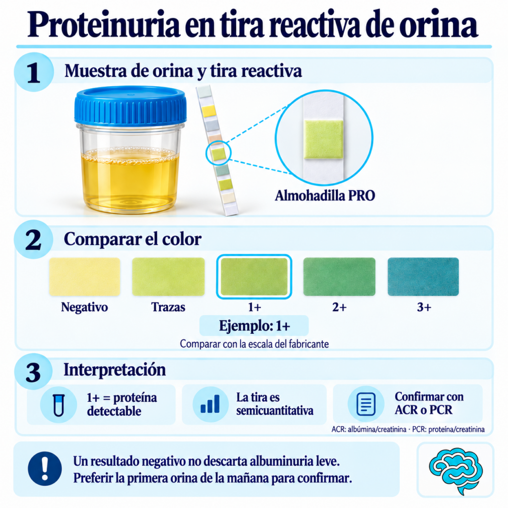
Image 3 – Rapid urine test showing proteinuria.
However, as we mentioned, the rapid test or urine colorimetry does not accurately measure how many proteins are excreted daily. Therefore, a positive result should be confirmed with a quantitative test.
Theurine collection of 24 hours.is the reference method for measuring total protein excretion.
To perform it, the patient discards the first urination and notes the time. From that moment on, they collectallthe urine during the day and night; upon reaching the same time the next day, they urinate one last time and include that sample.
The container indicated by the laboratory is used andit is usually not necessary to place a urinary catheter. If any urination is missed, the result may be inaccurate.
However,Nephrotic range proteinuria does not automatically mean nephrotic syndrome..
Let us remember that the normal value of serum albumin is 3,4–5,4 g/dL. Therefore, we are talking about hypoalbuminemia when we have a patient with less than 3,4 g/dL.
However, in nephrotic syndrome, lower ranges are often observed, frequently finding values between 3,0 g/dL and even less than 2,5 g/dL.
Edema is a common manifestation and results from several mechanisms, including decreased oncotic pressure and renal retention of sodium and water.
The key then is:intense proteinuria + hypoalbuminemia → nephrotic syndrome.
Super Pro Tip ✨: To remember it easily, just learn 3,5. Proteins above 3,5, Albumin below 3,5. Both are nephrotic syndrome
Finally,Diagnosing the syndrome is just the first step..
Next, it is necessary to investigate what disease is causing the glomerular damage. The medical history, physical examination, and tests guide this search. In certain cases, arenal biopsyis needed to identify the disease and decide on treatment.
Look for the cause, do not stop at the syndrome.
The medical history reviews diabetes, autoimmune diseases, infections, medications, family history, and systemic symptoms.
Based on that suspicion, targeted tests are requested: for example, glucose or glycosylated hemoglobin, complements, serologies for hepatitis and HIV, autoimmune studies, or anti-PLA2R antibodies. Requesting all tests from all patients without a clinical hypothesis does not necessarily improve reasoning.
Theanti-PLA2R antibodiesareautoantibodies: defenses that mistakenly recognize a protein of the body itself.PLA2Rmeanstype M phospholipase A₂ receptorand is found in thepodocytes. A positive result, in a person with nephrotic syndrome, suggests membranous nephropathy associated with PLA2R. However, a negative result does not rule out membranous nephropathy, especially in early stages.
In adults, a kidney biopsy is usually considered to define the lesion and guide treatment, although there are exceptions. KDIGO indicates that membranous nephropathy can be diagnosed without a biopsy in a patient with nephrotic syndrome and positive anti-PLA2R antibodies, always within a complete clinical assessment.
In children with typical presentation, an initial biopsy is generally not necessary; it is reserved for treatment resistance or atypical features such as macroscopic hematuria, low complement, sustained hypertension, or acute kidney injury that cannot be explained by hypovolemia.
An important challenge is distinguishing nephrotic syndrome from nephritic syndromeThis last one is characterized by glomerular inflammation, hematuria, and often hypertension and reduced filtration.
Both patterns can overlap; a person with significant proteinuria may also have hematuria. The comparison guides but does not replace the study of the cause.
Algorithm of clinical correlation and pathophysiology of nephrotic syndrome
Below we present a summary of the pathophysiology of nephrotic syndrome and its respective clinical correlation:
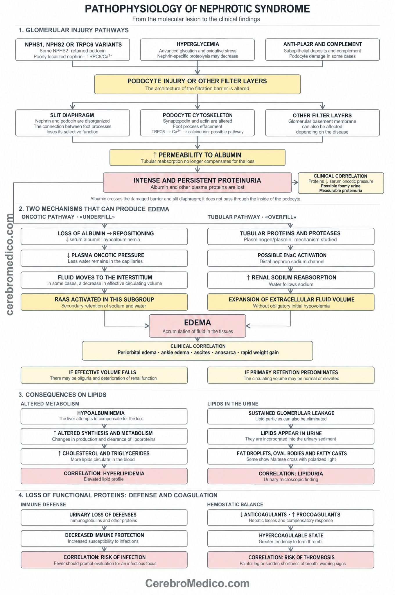
Image 4 – Algorithm of clinical pathophysiological correlation of nephrotic syndrome
Treatment of nephrotic syndrome
The treatment has two simultaneous objectives; to control the consequences of proteinuria and to act on the disease that produces it.
It is important to remember that treatment is not just diuretics, as administering only this may leave the filter still damaged; if we only address the cause, edema or a thrombotic complication may require immediate measures.
Support measures and prevention of complications
Reducing sodium in the diet helps limit water retention. Diuretics are adjusted according to the intensity of edema, blood pressure, and renal function; an excess may reduce effective circulating volume and worsen renal function.
In appropriate patients, angiotensin-converting enzyme inhibitors (ACE inhibitors) or angiotensin receptor antagonists (ARBs) reduce blood pressure and proteinuria, with monitoring of potassium and creatinine.
Persistent hyperlipidemia is assessed according to cardiovascular risk, not just by an isolated figure during a brief episode.
Recommended vaccinations and early attention to infections are part of care. Preventive anticoagulation requires estimating the risk of thrombosis and bleeding, especially in membranous nephropathy or marked hypoalbuminemia; it is not automatically indicated for everyone with nephrotic syndrome.
Treatment of the underlying disease
In minimal change disease sensitive to steroids, glucocorticoids are a central part of the treatment.
The KDIGO pediatric guideline 2025 recommends initial courses of 8 or 12 weeks in children with newly diagnosed nephrotic syndrome, with daily regimens and then on alternate days; frequent relapses or resistance necessitate reconsidering the strategy and sometimes using steroid-sparing drugs.
These decisions are individualized based on age, progression, and adverse effects.
In adults with minimal change or primary FSGS, treatment may include immunosuppression guided by the lesion and response.
In secondary FSGS, the cause is treated and renal protective measures are optimized; it should not be assumed that it will respond to the same drugs as the primary form.
Membranous nephropathy requires risk stratification and monitoring of proteinuria, renal function, and, when appropriate, anti-PLA2R before deciding on immunosuppression.
Diabetes, lupus, infections, and amyloidosis also require specific management of each disease.
Follow-up measures weight, blood pressure, edema, proteinuria, albumin, creatinine, and possible adverse effects.
The remission of proteinuria is important because it is usually accompanied by recovery of albumin and a lower risk of complications. but the time to achieve it depends on the cause. Therefore, the prognosis is better explained by the etiological diagnosis and evolution than by the label “nephrotic syndrome” alone.
Aspectos clave de la fisiopatología del síndrome nefrótico
- – Damage to podocytes and their slit diaphragm—or other layers of the filter—alters the glomerular barrier and allows albumin to pass into the urine.
- – If the filtered albumin exceeds the tubular recovery capacity, persistent proteinuria appears. When the loss exceeds hepatic replenishment, serum albumin also decreases.
- – Nephrotic Syndrome should be considered whenever a patient has proteinuria greater than 3.5 g/dL and albuminemia less than 3,5 g/dL (usually between 3,0 and 2,5 g/dL). ⚕️
- – Hypoalbuminemia reduces oncotic pressure and promotes the movement of fluid into the interstitium. If the effective circulating volume falls, the RAAS is activated and sodium and water are retained (underfill).
- – Filtered proteins and proteases may promote the activation of ENaC and primary renal sodium retention (overfill). Edema does not always imply hypovolemia; both pathways can coexist.
- – The hepatic response alters lipoprotein metabolism and may elevate cholesterol and triglycerides. The excretion of lipids in urine produces lipiduria, visible as fatty bodies or casts.
- – The loss of defense proteins increases the risk of infections. The loss of anticoagulants and the procoagulant hepatic response favor thrombosis; the risk varies among patients.
- – Edema may begin in the eyelids or legs and be accompanied by rapid weight gain; foamy urine suggests but does not confirm proteinuria. Fever, sudden dyspnea, or painful unilateral swelling necessitate the search for complications.
Referencias consultadas23 fuentes consultadas
- Kidney Disease: Improving Global Outcomes (KDIGO) Nephrotic Syndrome in Children Work Group. KDIGO 2025 Clinical Practice Guideline for the Management of Nephrotic Syndrome in Children. Kidney Int. 2025;107(5S):S241-S289. doi:10.1016/j.kint.2024.11.007.
- National Institute of Diabetes and Digestive and Kidney Diseases. Nephrotic syndrome in adults [Internet]. Bethesda (MD): NIDDK; 2020 [consultado el 26 sep 2026]3. Vestergaard SV, Birn H, Jensen SK, Sørensen HT, Nitsch D, Christiansen CF. Twenty-four-year trends in incidence and mortality of nephrotic syndrome: a population-based cohort study. Epidemiology. 2023;34(3):411-420. doi:10.1097/EDE.0000000000001576.
- Lionaki S, Derebail VK, Hogan SL, Barbour S, Lee T, Hladunewich M, et al. Venous thromboembolism in patients with membranous nephropathy. Clin J Am Soc Nephrol. 2012;7(1):43-51. doi:10.2215/CJN.04250511.
- Chen JL, Wang L, Yao XM, Zang YJ, Wang Y, Li ZJ, et al. Association of urinary plasminogen-plasmin with edema and epithelial sodium channel activation in patients with nephrotic syndrome. Am J Nephrol. 2019;50(2):92-104. doi:10.1159/000501059.
- Svenningsen P, Bistrup C, Friis UG, Bertog M, Haerteis S, Krueger B, et al. Plasmin in nephrotic urine activates the epithelial sodium channel. J Am Soc Nephrol. 2009;20(2):299-310. doi:10.1681/ASN.2008040364.
- Stoycheff N, Stevens LA, Schmid CH, Tighiouart H, Lewis J, Atkins RC, et al. Nephrotic syndrome in diabetic kidney disease: an evaluation and update of the definition. Am J Kidney Dis. 2009;54(5):840-849. doi:10.1053/j.ajkd.2009.04.016.
- Kestilä M, Lenkkeri U, Männikkö M, Lamerdin J, McCready P, Putaala H, et al. Positionally cloned gene for a novel glomerular protein—nephrin—is mutated in congenital nephrotic syndrome. Mol Cell. 1998;1(4):575-582. doi:10.1016/S1097-2765(00)80057-X.
- Boute N, Gribouval O, Roselli S, Benessy F, Lee H, Fuchshuber A, et al. NPHS2, encoding the glomerular protein podocin, is mutated in autosomal recessive steroid-resistant nephrotic syndrome. Nat Genet. 2000;24(4):349-354. doi:10.1038/74166.
- Huber TB, Köttgen M, Schilling B, Walz G, Benzing T. Interaction with podocin facilitates nephrin signaling. J Biol Chem. 2001;276(45):41543-41546. doi:10.1074/jbc.C100452200.
- Verma R, Kovari I, Soofi A, Nihalani D, Patrie K, Holzman LB. Nephrin ectodomain engagement results in Src kinase activation, nephrin phosphorylation, Nck recruitment, and actin polymerization. J Clin Invest. 2006;116(5):1346-1359. doi:10.1172/JCI27414.
- Faul C, Donnelly M, Merscher-Gomez S, Chang YH, Franz S, Delfgaauw J, et al. The actin cytoskeleton of kidney podocytes is a direct target of the antiproteinuric effect of cyclosporine A. Nat Med. 2008;14(9):931-938. doi:10.1038/nm.1857.
- Verheijden KAT, Sonneveld R, Bakker-van Bebber M, Wetzels JFM, van der Vlag J, Nijenhuis T. The calcium-dependent protease calpain-1 links TRPC6 activity to podocyte injury. J Am Soc Nephrol. 2018;29(8):2099-2109. doi:10.1681/ASN.2016111248.
- Beck LH Jr, Bonegio RGB, Lambeau G, Beck DM, Powell DW, Cummins TD, et al. M-type phospholipase A2 receptor as target antigen in idiopathic membranous nephropathy. N Engl J Med. 2009;361(1):11-21. doi:10.1056/NEJMoa0810457.
- Haddad G, Lorenzen JM, Ma H, de Haan N, Seeger H, Zaghrini C, et al. Altered glycosylation of IgG4 promotes lectin complement pathway activation in anti-PLA2R1-associated membranous nephropathy. J Clin Invest. 2021;131(5):e140453. doi:10.1172/JCI140453.
- Vivarelli M, Massella L, Ruggiero B, Emma F. Minimal change disease. Clin J Am Soc Nephrol. 2017;12(2):332-345. doi:10.2215/CJN.05000516.
- Singh A, Fridén V, Dasgupta I, Foster RR, Welsh GI, Tooke JE, et al. High glucose causes dysfunction of the human glomerular endothelial glycocalyx. Am J Physiol Renal Physiol. 2011;300(1):F40-F48. doi:10.1152/ajprenal.00103.2010.
- Chen YM, Kikkawa Y, Miner JH. A missense LAMB2 mutation causes congenital nephrotic syndrome by impairing laminin secretion. J Am Soc Nephrol. 2011;22(5):849-858. doi:10.1681/ASN.2010060632.
- Ray EC, Rondon-Berrios H, Boyd CR, Kleyman TR. Sodium retention and volume expansion in nephrotic syndrome: implications for hypertension. Adv Chronic Kidney Dis. 2015;22(3):179-184. doi:10.1053/j.ackd.2014.11.006.
- Agrawal S, Zaritsky JJ, Fornoni A, Smoyer WE. Dyslipidaemia in nephrotic syndrome: mechanisms and treatment. Nat Rev Nephrol. 2018;14(1):57-70. doi:10.1038/nrneph.2017.155.
- Haas ME, Levenson AE, Sun X, Liao WH, Rutkowski JM, de Ferranti SD, et al. The role of proprotein convertase subtilisin/kexin type 9 in nephrotic syndrome-associated hypercholesterolemia. Circulation. 2016;134(1):61-72. doi:10.1161/CIRCULATIONAHA.115.020912.
- Abdelghani E, Waller AP, Wolfgang KJ, Stanek JR, Parikh SV, Rovin BH, et al. Exploring the role of antithrombin in nephrotic syndrome-associated hypercoagulopathy: a multi-cohort study and meta-analysis. Clin J Am Soc Nephrol. 2023;18(2):234-244. doi:10.2215/CJN.0000000000000047.
- Schmidt-Gayk H, Grawunder C, Tschöpe W, Schmitt W, Ritz E, Pietsch V, et al. 25-hydroxy-vitamin-D in nephrotic syndrome. Lancet. 1977;2(8029):105-108. doi:10.1016/S0140-6736(77)90118-0.
- Matsell DG, Wyatt RJ. The role of I and B in peritonitis associated with the nephrotic syndrome of childhood. Pediatr Res. 1993;34(1):84-88. doi:10.1203/00006450-199307000-00019.
¿Te ayudó este artículo?
Dale un cerebro para apoyar este contenido médico.


¿Qué te pareció este artículo?
Si te quedó alguna duda, déjala aquí. También puedes compartir tu experiencia.
Inicia sesión o crea tu cuenta en un par de clics. Completarás tu perfil solo cuando uses CerebroMédico IA.
Todavía no hay comentarios. Puedes abrir la conversación.